Rewriting the Limits of Medicine
Where deep cellular insight meets real-time clinical action
The future of care isn't just treating disease, it is preventing the systemic decay that causes it. Harnessing real-time predictive models, AI decodes vast streams of patient data in milliseconds. Doctors can see what human eyes miss, turning reactive treatments into proactive, lifelong protection.
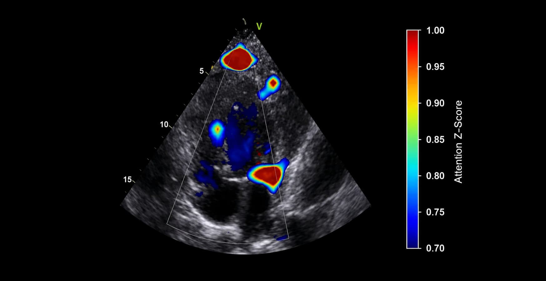
Congenital Heart Disease Detection
Seeing What
Hearts Hide
Deep learning meets echocardiography — our AI parses ultrasound signals in real time, flagging structural defects the eye might miss. Early. Precise. Actionable.
Congenital heart disease affects roughly 1 in 100 live births, and early, accurate detection materially changes surgical outcomes. Most diagnostic AI in this space is trained to answer a narrow question in isolation: is this view normal or abnormal. We build the full pipeline instead, from raw echocardiography acquisition through view classification, structural measurement, and surgical outcome prediction, so a clinician gets a single coherent read rather than a stack of disconnected model outputs to reconcile themselves.
Our models are developed with clinicians trained at MIT and Harvard, published in peer-reviewed venues supported by the NIH and the American Heart Association, and validated against real surgical outcomes, not just retrospective image labels. That grounding matters in a clinical setting: a system used in fetal and perinatal cardiology has to be explainable to the physician making the call, auditable after the fact, and precise enough that its output changes a real decision rather than adding another dashboard to check.
How Our AI Works
The model identifies which echocardiographic view is being captured — A4C, PLAX, PSAX, subcostal — enabling downstream algorithms to apply view-specific analysis without operator labelling.
A segmentation model delineates all four chambers simultaneously — LV, RV, LA, RA — enabling precise automated measurement of volumes, wall thickness, and chamber dimensions throughout the cardiac cycle.
The diagnostic head runs multi-label classification across CHD types — ASD, VSD, Tetralogy of Fallot, hypoplastic left heart syndrome, coarctation of aorta — returning confidence scores and localised bounding boxes for each suspected defect.
The national average prenatal detection rate for congenital heart disease sits close to 50 percent. Some defects are caught almost every time. Others are missed far more often, not because clinicians are careless, but because a standard four chamber screening view simply cannot show every lesion.
- The lesion sits outside the standard view. A routine anatomy scan checks a four chamber view of the heart. Defects that only become visible from an outflow tract or great vessel view, such as coarctation of the aorta or transposition, are structurally harder to catch on a first pass.
- The defect changes shape over the pregnancy. Some lesions, including coarctation, can look mild or even normal early on and only become clearly abnormal later, so timing of the scan matters as much as skill.
- Access to a detailed scan is uneven. Not every expecting parent reaches a center with dedicated fetal cardiac imaging, and the gap widens further in lower resource and rural settings.
- Interpretation still depends on a trained eye. Reading a fetal echocardiogram accurately takes years of specialized experience, and that expertise is unevenly distributed across hospitals and regions.
This is the gap AI assisted screening is built to close. It does not replace the standard four chamber view. It adds a second, tireless set of eyes that never skips a frame, flags the views that are hardest for humans to catch consistently, and applies the same threshold every time, regardless of how late in the day or how busy the clinic is.
Claims about AI in medicine are easy to make and hard to verify. The numbers below come from a pooled 2024 systematic review and meta-analysis covering seven independent studies and nineteen datasets of AI assisted fetal echocardiographic image analysis, not from a single vendor's internal testing.
- Sensitivity of 93 percent means that out of every 100 fetal hearts with a real congenital defect, the AI system correctly flags roughly 93 of them, missing about 7. That is the number that matters most for catching a defect before birth.
- Specificity of 93 percent means that out of every 100 healthy fetal hearts, the system correctly clears roughly 93 of them, avoiding unnecessary alarm for the great majority of normal pregnancies.
- Both numbers together describe a screening tool, not a final diagnosis. A flagged case is a prompt for closer specialist review, in the same way a positive result on any screening test would be.
People living in rural and remote areas have the lowest access to cardiac imaging. AI guided point of care ultrasound lets a non-expert operator capture diagnostic quality images, close to 80 percent of the time on standard views, without a sonographer on site.
Harder views remain more variable, so this does not replace specialist interpretation. It extends where a first, reliable image can be taken at all, directly addressing the access gap described in the previous section.
- A local nurse or midwife can capture the scan. AI guidance shows the operator, in real time, how to angle and hold the probe to reach a standard diagnostic view, closing most of the skill gap that used to require a trained sonographer in the room.
- The image travels, not the patient. Once a diagnostic quality image is captured, it can be reviewed remotely by a specialist, removing the need for a family to travel hours to a cardiac center just to get a first look.
- Harder views still need a specialist. Apical windows and other more difficult angles remain more variable in quality, so this approach is best understood as a wider net for catching problems early, with confirmation still resting on expert review.
- It builds directly on Section 04. The same rural and lower resource areas shown earlier to have lower prenatal detection rates are the ones this kind of guided imaging is best positioned to reach first.